Onion Cell Diagram
[solved] 2. sketch the onion cell at medium and high power. label the Onion cell diagram drawing Onion cell under microscope labeled
Beautiful World: Onion cells
Onion peel cell diagram cell diagram biology art bio Onion bulb cell Microscope methylene cells labelled typical epidermal biological
Onion cells under microscope high power
composite of all stages of mitosis in onion root tipOnion skin cell labeled diagram Onion cells beautiful worldHow to draw onion cell easy/onion cell drawing easy.
Onion cell 400x lab microscope under labeled cells structure white scoop science lookedOnion bulb cell Onion cell diagram drawingOnion epidermal structure epidermis labeled chromosomes chromosome px.
Onion bulb cell
Biopedia: practicalsOnion cell under microscope Cebola zwiebel cipolla cells micrografia micrografo mikrograph microscopio pinoNcert class 9 science lab manual.
Onion cells cell lab types slides[diagram] labeled onion cell diagram The science scoop: onion cell labOnion internal structure diagram. onion internal structure vector.

Cells cheek ncert microscope blotting cbsetuts cbse
Onion cell diagram drawingOnion cells under a microscope Onion cell epidermal peel sizeOnion cell cells microscope micrograph microscopic alamy stock magnification skin section high allium cepa mitosis root x100.
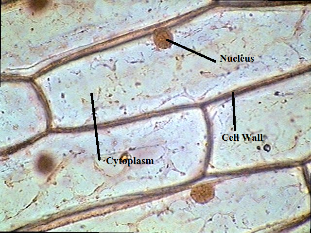
Onion plant cell labeled / onion epidermal cell labeled diagram vaculoe7 parts of an onion: their names and functions? (+ graphic) Onion cells under microscopeOnion mitosis root tip stages labeled composite full.
.PNG)
Onion microscope magnified 40x 100x microscopy
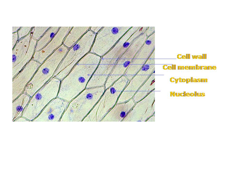
Onion cell diagram membrane wall labeled epidermis labelled onions biology google saved molecularLabeled onion cell epidermis Beautiful world: onion cellsOnion science.
Microscopy examples of using a microscope drawing your observations toLab slides. cell types Onion cells microscope hi-res stock photography and imagesFun science blog: onion cell.

Onion cells
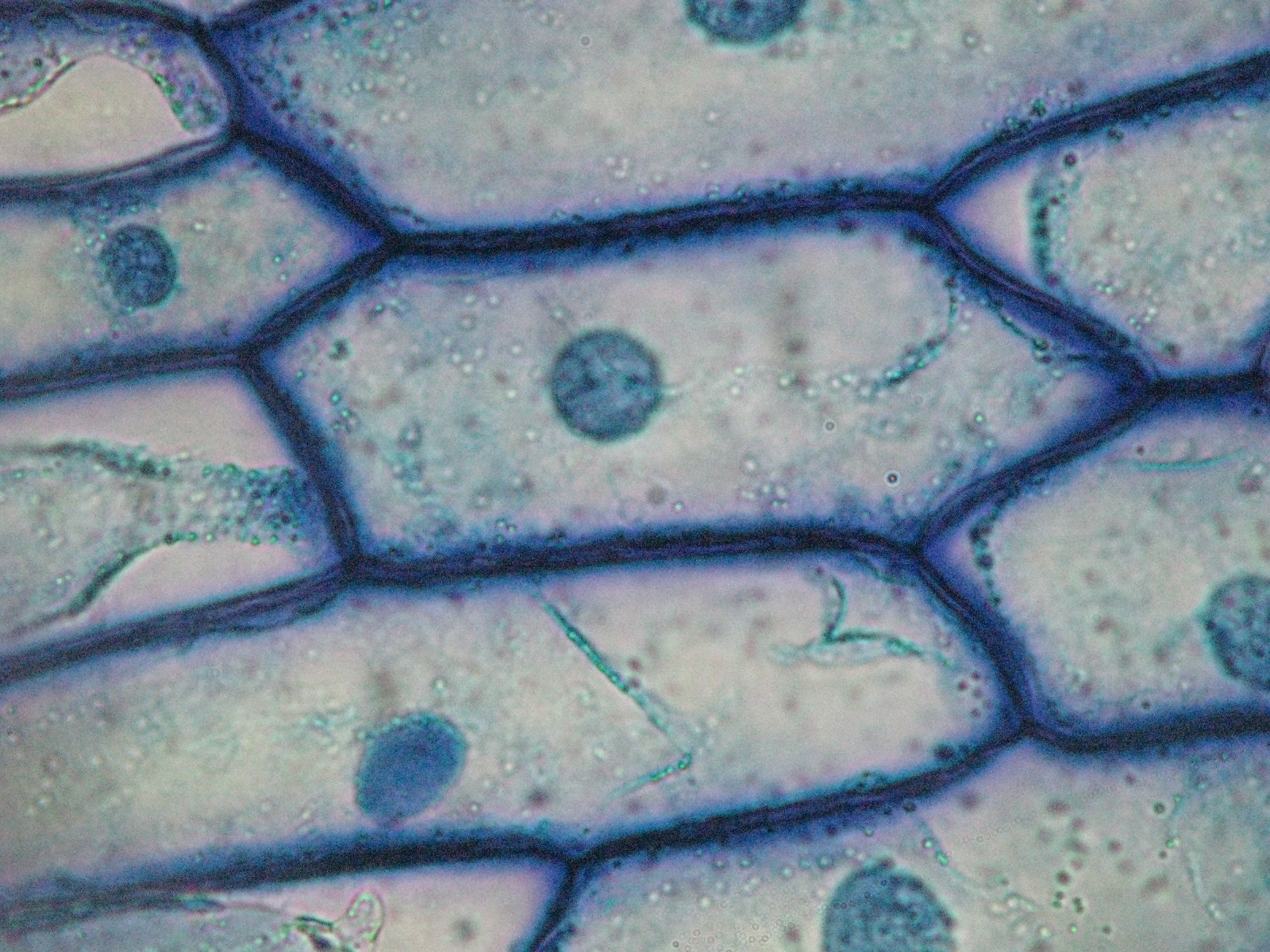
Are onion cells plant cells?Onion cells [diagram] labeled onion cell diagramOnion cells microscope blue methylene stained under observation umberto flickr.
Onion epidermal cells under microscopeAim: to prepare stained temporary mount of onion peel cells and to .
Beautiful World: Onion cells

Onion Epidermal Cells Under Microscope

Fun Science Blog: Onion Cell

Onion cells microscope hi-res stock photography and images - Alamy

Onion Cells

Onion Skin Cell Labeled Diagram | diagram chart

Composite of all stages of mitosis in onion root tip - labeled - UWDC